
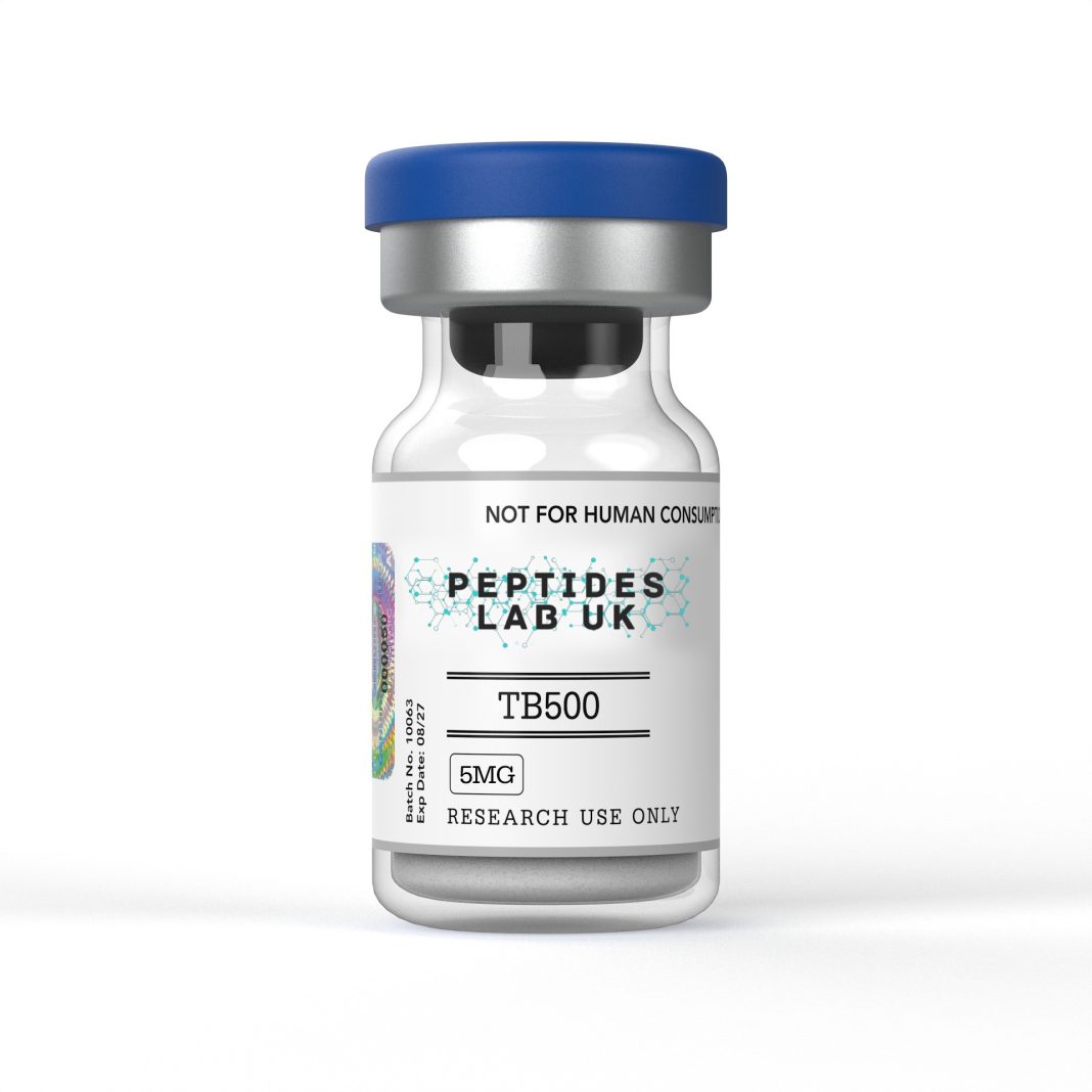
TB-500 is a synthetic peptide corresponding to the LKKTETQ active domain of Thymosin Beta-4, a 43-amino-acid protein and the principal G-actin-sequestering molecule in mammalian cells. The synthetic fragment consists of the acetylated seven-amino-acid sequence Ac-LKKTETQ, representing residues 17 to 23 of the full-length parent protein, with a molecular weight of approximately 889 Da, considerably lower than the roughly 4,963 Da of full-length Thymosin Beta-4. Within laboratory research, TB-500 peptide is used as a tool compound for studying actin cytoskeletal dynamics, cell migration and angiogenesis-related signalling pathways associated with the parent protein’s actin-binding domain.
What Is TB-500 Peptide?
TB-500 is a synthetic derivative representing a specific functional fragment of Thymosin Beta-4 (Tβ4), one of the most abundant and highly conserved polypeptides found across mammalian cells and tissues. Full-length Tβ4 is a 43-amino-acid protein that functions as the primary intracellular G-actin sequestering protein, meaning it binds monomeric actin and regulates the pool available for polymerisation into filamentous actin (F-actin), a process fundamental to cell shape, motility and migration. TB-500 corresponds specifically to the 17-amino-acid region of Tβ4 that has been identified as its principal actin-binding motif, centred on the LKKTETQ sequence spanning residues 17 to 23.
This structural relationship is an important distinction for researchers to understand, since the great majority of the published efficacy literature on wound healing, cardiac repair and corneal healing was generated using the full-length 43-amino-acid Tβ4 protein rather than the isolated LKKTETQ fragment. The fragment retains the core actin-binding motif and, in in-vitro assays, has been shown to reproduce aspects of the parent protein’s actin-sequestering activity, but researchers should treat findings generated with full-length Tβ4 and findings generated with the isolated TB-500 fragment as related but distinct bodies of evidence rather than directly interchangeable.
A further structural feature relevant to TB-500’s research profile is its low molecular weight and small size relative to the full-length protein, which has been associated in the literature with greater capacity for diffusion and systemic dispersion across tissue compartments in animal-model studies, a property proposed to contribute to reports of activity at sites distant from a local injection point in preclinical wound-healing and cardiac-repair research. Structural analyses using X-ray crystallography of the actin-binding interface have confirmed that this region of the Tβ4 sequence, which includes the LKKTETQ motif, forms a one-to-one complex with G-actin, sequestering the monomer by capping both ends of the actin structure and preventing its incorporation into growing filaments.
Mechanism of Action
The central and best-characterised mechanism associated with the TB-500 active domain is sequestration of monomeric G-actin. Thymosin Beta-4, and by extension the LKKTETQ fragment that constitutes TB-500, binds G-actin with high affinity, maintaining a portion of the total cellular actin pool in a polymerisation-incompetent state. Structural work resolving the gelsolin-domain-1-thymosin-beta-4 hybrid bound to actin at high resolution has confirmed that this binding involves the actin-interacting WH2 motif, capping both ends of the actin monomer and preventing nucleotide exchange, which keeps actin sequestered until cellular signalling triggers its release for polymerisation.
This actin-sequestering activity underlies the peptide’s proposed downstream effects on cell migration. By regulating the pool of free G-actin available for polymerisation, the LKKTETQ domain is understood to influence the formation of lamellipodia and filopodia, the actin-rich structures at the leading edge of a migrating cell that drive directional movement through tissue. This mechanism is the proposed basis for reported effects on fibroblast, keratinocyte and endothelial cell migration observed in cell-culture wound-healing assays.
Angiogenesis represents a second major mechanistic focus in the literature. Research examining the actin-binding site on Thymosin Beta-4 directly has reported that this same seven-amino-acid region functions as the major cell-adhesion site on the full-length molecule, and that adhesion mediated through this site could be blocked using the isolated seven-amino-acid peptide, demonstrating its role as an essential component of the protein’s angiogenic activity in endothelial cell and aortic ring sprouting assays. Reports in the literature associate this angiogenic activity with upregulation of vascular endothelial growth factor (VEGF) signalling, promoting endothelial cell migration, adhesion and tubule formation, key steps in the formation of new vascular structures studied in preclinical angiogenesis models.
A further mechanistic pathway relevant to tissue-repair research is Thymosin Beta-4’s reported interaction with laminin-5, a component of the extracellular matrix and basement membrane involved in epithelial cell adhesion and migration. This interaction has been proposed as a contributing mechanism in corneal and dermal epithelial wound-healing research, where restoration of an intact epithelial layer depends on coordinated keratinocyte or corneal epithelial cell migration across the basement membrane. Anti-inflammatory pathway modulation has also been documented for the full-length protein, including reported suppression of pro-inflammatory cytokine signalling such as interleukin-8 production following stimulation by tumour necrosis factor-alpha, alongside down-regulation of nuclear factor-kappa B (NF-κB) activity, a transcription factor central to inflammatory gene expression, in cell-culture models of inflammatory signalling.
What the Research Shows
Foundational structural and mechanistic characterisation of the actin-binding domain was reported in a study examining the specific seven-amino-acid actin-binding site on Thymosin Beta-4, which found that this motif was the major cell-adhesion site on the molecule and demonstrated that adhesion mediated through this site was blocked by the same isolated peptide fragment, confirming the motif’s essential role in angiogenic activity in endothelial and aortic ring assay models (actin-binding site angiogenesis study).
A regulatory and analytical study addressing TB-500 specifically, rather than the full-length parent protein, examined its use as a veterinary preparation and developed a liquid chromatography-mass spectrometry method to detect the synthetic LKKTETQ peptide with artificial N-terminal acetylation in equine urine and plasma, confirming the compound’s specific molecular identity as distinct from naturally occurring Thymosin Beta-4 fragments (TB-500 analytical detection study).
Corneal epithelial research has focused primarily on full-length Thymosin Beta-4, with a review describing its properties as a corneal wound-healing and anti-inflammatory agent, summarising cell-culture and animal-model evidence for accelerated corneal epithelial cell migration and reduced inflammatory markers following Tβ4 application in experimental corneal injury models (corneal wound healing review).
Cardiac tissue repair research has similarly relied primarily on full-length Tβ4 administered in rodent models of myocardial infarction. Studies using permanent ligation of the left anterior descending coronary artery in mice have reported improved ejection fraction and reduced infarct scar volume following Tβ4 treatment initiated shortly after injury, with proposed mechanisms including preservation of ischemic cardiomyocytes through pro-survival signalling and subsequent stimulation of coronary blood vessel growth, findings that researchers have proposed operate through both an acute anti-apoptotic phase and a later phase involving vascular and cardiac progenitor cell activation. Broader reviews of Thymosin Beta-4’s structural and functional properties have further catalogued reported effects across cell migration, blood vessel formation, cell survival and modulation of cytokine and matrix-remodelling gene expression, forming the basis for continued dermal, corneal and cardiac preclinical research programmes.
Researchers should note throughout this body of evidence that the isolated LKKTETQ fragment corresponding to TB-500 has been tested directly in comparatively few of these specific efficacy models, with much of the published cardiac, corneal and dermal literature generated using the full 43-amino-acid Tβ4 protein rather than the fragment alone.
Research Applications
Within laboratory settings, TB-500 peptide and its parent domain are used across several established research contexts. Endothelial cell tubulogenesis assays represent a common application, in which researchers examine tube-formation capacity in cultured endothelial cells following exposure to the actin-binding peptide, using this in-vitro model as a proxy for angiogenic potential relevant to the VEGF-associated signalling described in the mechanism section. Fibroblast migration protocols, typically using scratch-wound or transwell migration assay formats, are used to quantify directional cell movement in response to peptide exposure, providing a functional readout linked to the actin-sequestration mechanism.
Extracellular matrix remodelling research constitutes a further application area, examining how actin cytoskeletal reorganisation driven by G-actin sequestration intersects with matrix protein expression and basement membrane interactions, including the laminin-5 interaction relevant to epithelial wound-healing research. Tissue engineering setups have also incorporated TB-500 and related Thymosin Beta-4 fragments as candidate signalling components in scaffold-based cell culture systems, where researchers investigate whether actin-modulating peptides can influence cell seeding, migration and organisation within engineered tissue constructs. When selecting a certified TB-500 research peptide for endothelial migration or cell assay protocols, researchers should confirm the exact acetylated sequence and purity documentation supplied, since the artificial N-terminal acetylation characteristic of TB-500 distinguishes it analytically from naturally occurring Thymosin Beta-4 fragments and is relevant to assay reproducibility.
Comparative research has also examined engineered tandem variants combining two TB4 domains, reporting that such constructs can sequester a larger pool of G-actin than the single fragment, providing researchers with a broader comparative framework for studying dose-dependent actin-sequestration effects across different peptide formats in cell-culture and animal-model systems.
Purity, Storage and Handling
Research-grade TB-500 should be accompanied by a certificate of analysis confirming purity by HPLC, typically at or above 98 percent, together with mass spectrometry verification confirming the correct acetylated LKKTETQ sequence and molecular weight of approximately 889 Da. Because TB-500 is a synthetic fragment distinct from naturally occurring Thymosin Beta-4, analytical confirmation of both sequence accuracy and the artificial N-terminal acetylation is particularly relevant when comparing findings against the published fragment-specific literature rather than full-length Tβ4 studies. When evaluating high-purity TB-500 peptide for laboratory research, UK researchers should verify that each batch includes this documentation rather than relying on a generic product listing.
Lyophilised TB-500 should be stored at -20°C, protected from light and moisture, in order to preserve peptide integrity prior to reconstitution. Once reconstituted, the peptide should be handled promptly and kept refrigerated at 2-8°C, since reconstituted peptide solutions are generally more vulnerable to degradation through oxidation and repeated freeze-thaw cycling than the lyophilised form. Aliquoting reconstituted material into single-use volumes is recommended to minimise freeze-thaw exposure across an experimental run, and researchers should follow the supplier’s stated stability window for reconstituted material rather than relying on visual inspection to assess degradation.
Frequently Asked Questions
How does TB-500 differ structurally from full-length Thymosin Beta-4?
TB-500 corresponds to the Ac-LKKTETQ sequence, residues 17 to 23 of the 43-amino-acid Thymosin Beta-4 protein, with a molecular weight of approximately 889 Da compared with roughly 4,963 Da for the full-length parent molecule. The fragment retains the principal actin-binding motif but does not include the complete Tβ4 sequence used in most published efficacy studies.
What is the primary mechanism attributed to the TB-500 active domain?
The LKKTETQ sequence is understood to sequester monomeric G-actin, regulating the pool of actin available for polymerisation into filamentous actin. This actin-binding activity is proposed as the mechanistic basis for reported effects on cell migration and angiogenic signalling documented in the broader Thymosin Beta-4 literature.
Is most of the published TB-500 research conducted using the fragment itself?
No. The majority of published wound-healing, cardiac-repair and corneal-healing studies have used the full-length 43-amino-acid Thymosin Beta-4 protein rather than the isolated LKKTETQ fragment. Researchers should distinguish fragment-specific findings, such as analytical detection studies, from the broader full-length protein literature when interpreting research on TB-500 specifically.
How should research-grade TB-500 be verified before use in an assay?
Researchers should request a batch-specific certificate of analysis confirming HPLC purity of 98 percent or higher and mass spectrometry confirmation of the correct acetylated sequence, since both sequence accuracy and the artificial acetylation status are relevant to reproducing actin-sequestration and cell-migration assay findings.
TB-500 peptide, as supplied by Peptides Lab UK and comparable UK research suppliers, is intended strictly for in-vitro and animal-model laboratory research. It is not licensed or intended for human or veterinary use, and nothing in this article should be interpreted as guidance for personal administration.